An Ophthalmoscope Is Used to Determine Which of the Following
Small spot large spot semicircle red-free filter and fixed. Shine the ophthalmoscope light into the patients pupil at arms length and observe the red reflex.
Fundoscopy Ophthalmoscopy Osce Guide Geeky Medics
Which of the following is a flexible a traumatic device used to facilitate proper placement of catheters into lumens of vessels ducts or ureters.

. The ophthalmoscope can also be used for examining the anterior part of the eye by turning the lens dial to 10. ABase the pace of the examination on the patients needs and abilities. Regardless of model type these hand-held devices are critical in the evaluation and diagnosis of a variety of diseases in the eye.
Retinal tear or detachment. The health care provider performs this exam by shining a beam of light through the pupil using an instrument called an ophthalmoscope. Verify doubtful papillary action c.
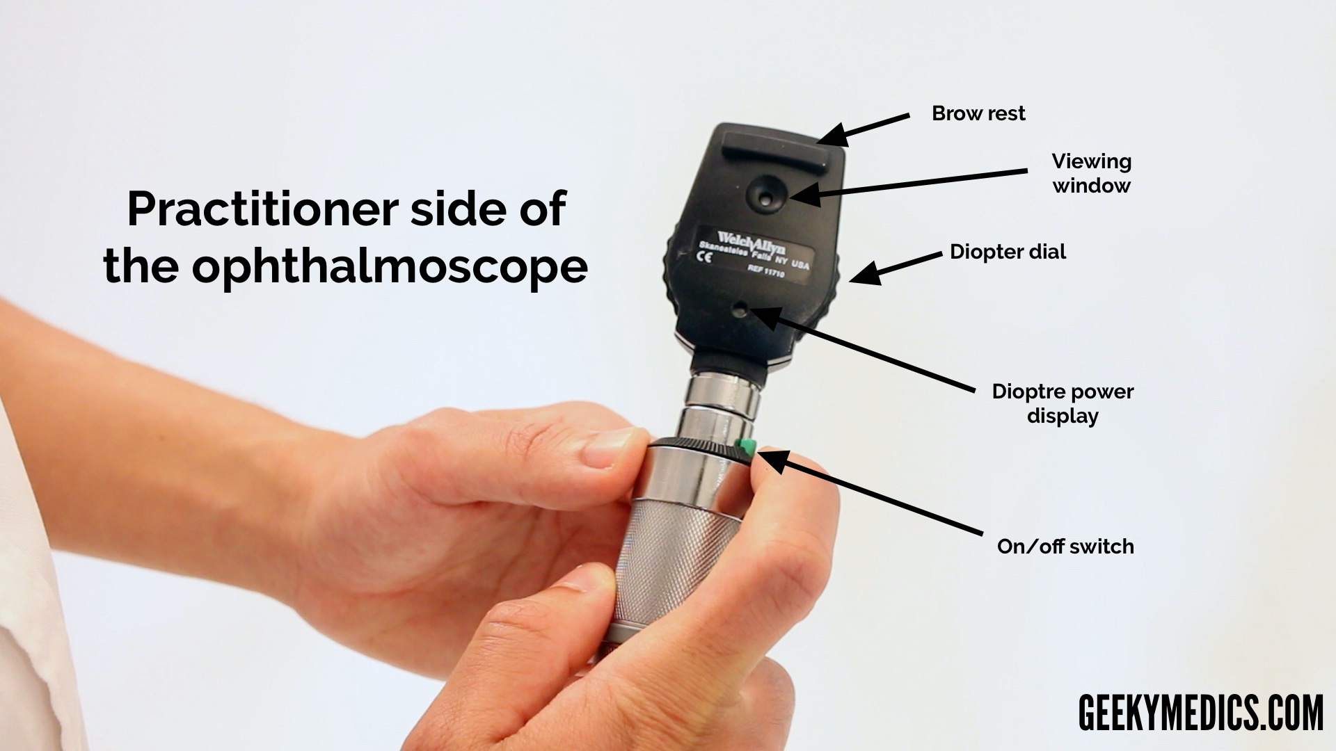
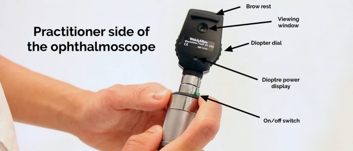
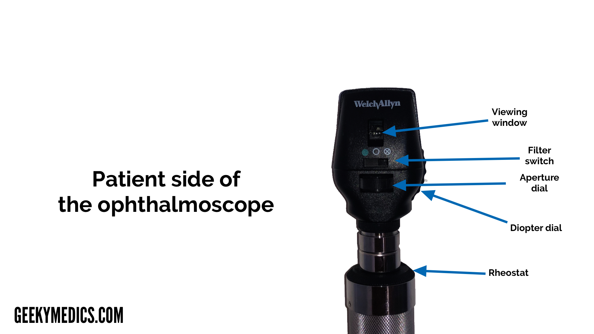
An ophthalmoscope is used for a funduscopic examination an examination of the internal structures of the eye. I have a question about the ophthalmoscope. The ophthalmoscope has 5 apertures.
The _____ uses ultrasound waves to determine the diopter size of the intraocular lens to insert following cataract extraction. In its full-blown form papilledema has all the manifestations of axoplasmic constipation and peripapillary venous obstruction Figure 70-1. It produces a printout of the shape of the eye as well as the optimal lens size for that patient.
Ophthalmoscopy also called fundoscopy or a fundoscopic exam is a common procedure performed by an eye doctor. Funduscopy with a direct ophthalmoscope or indirect slit-lamp ophthalmoscope remains the easiest and most sensitive means of detecting papilledema. To examine myopic patients you use the negative red numbers and to examine hyperopic patients you use the green positive numbers.
The exam involves the use of special lenses and bright direct light to provide a better. Optometrists and general practitioners alike can use an ophthalmoscope to diagnose or monitor diseases of the eye as well as conditions like hypertension and diabetes. Proper use of an ophthalmoscope requires a bit of practice and familiarity with the functions of your device.
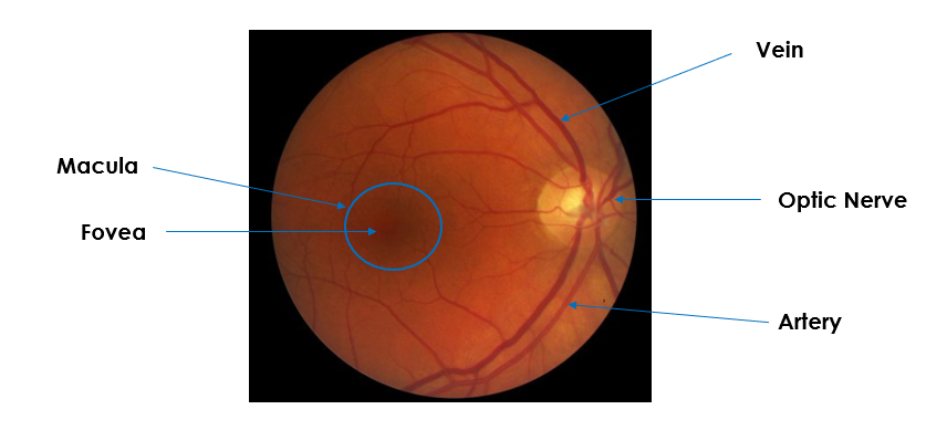
An ophthalmoscope is about the size of a flashlight. Pediatricians and general practitioners may also include ophthalmoscopy in routine physical exams. An ophthalmoscope is particularly useful for examining the structures of the retinathe light sensitive area at the back of the eye responsible for processing images.
Assessing skin with the use of pads of fingertips. They are used all over the world and are an essential piece of apparatus for all who wish to study the intricate biology of the eye. The lenses of the ophthalmoscope can be used to focus variously the apex and base of any intraocular mass and thus helps determine its height in dioptres.
Ophthalmoscopy also called fundoscopy is an exam your doctor optometrist or ophthalmologist uses to look into the back of your eyeWith it they can see the retina which senses light and. All of the above. The ophthalmoscope has 5 apertures which aperture.
Prime among these is the intended clinical role. An expected part of every eye exam ophthalmoscopes are capable of recognising healthy. Using the ophthalmoscope light as a pen light briefly examine the external features of the eye including lashes lid margins conjunctiva sclera iris and pupil shape size and reactivity.
Reviewed by Brian Boxer Wachler MD. An ophthalmoscope is a piece of equipment utilised by ophthalmologists that are used to inspect the internal structure of your eyes containing the retina. Red reflex anterior segment disc vessels and lastly macula see box.
Placing the patient in a supine position. Which aperture would be used to assess the eyes of a patient with undilated pupils C small at the conclusion of the examination the examiner shouldD summarize findings to the patient When the practitioner enters the examining room the infant patient is asleep. How to select an appropriate ophthalmoscope.
Ophthalmoscopy also called funduscopy is a test that allows a health professional to see inside the fundus of the eye and other structures using an ophthalmoscope or funduscopeIt is done as part of an eye examination and may be done as part of a routine physical examinationIt is crucial in determining the health of the retina optic disc and vitreous humor. Washing hands under warm water. There are two main types of.
If youve ever been for an eye test or visited an ophthalmologist theres a good chance they would have taken a look at your retina with an ophthalmoscope. The ophthalmoscope selection should be guided by a number of factors. You will be seated in a darkened room.
Upgrade to remove ads. The practitioner would best. What am I looking for.
It has a light and different tiny lenses that allow the provider to view the back of the eyeball. An ophthalmoscope is an instrument used to examine the retina. The ophthalmoscope can be used to.
These include hyperemia and elevation of the optic disc with obscuration of the. Which of the following is considered when preparing to examine an older adult. Your eye doctor can use ophthalmoscopy to screen for eye diseases and conditions that can affect blood vessels.
Detect foreign bodies in the cornea b. Traditionally part of almost every eye exam ophthalmoscopes can identify healthy structures within the eyeball and easily help your eye doctor see symptoms or indicators of diseases of the eye. You will either lie.
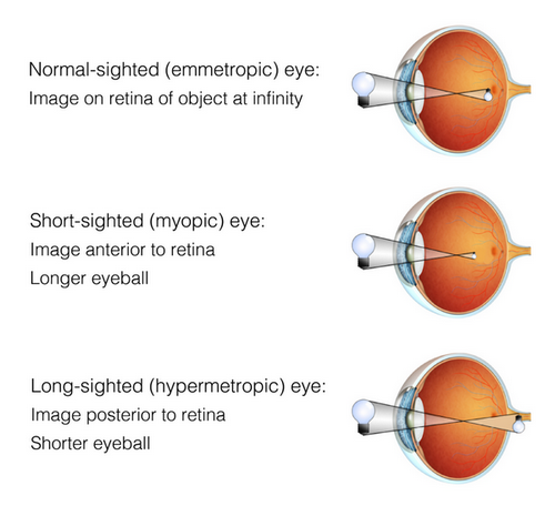
If youre farsighted looking into the eye of a nearsighted or myopic patient you need to concave the lens. Arrange the steps of palpation in the order the nurse would perform them during a patients physical examination. The ophthalmoscope also known as a fundoscope is a tool used in medicine to examine the interior of the eye including the retina fovea choroid macula optic disc and blood vessels.
I want to know how a doctor with an ametropic eye observes a patient. ADC Pocket Ophthalmoscopes have five aperture selections shown above. Pulmonary capillary wedge pressure.
Detect lens opacities d. Damage to your optic nerve.
Fundoscopy Ophthalmoscopy Osce Guide Geeky Medics

How To Use An Ophthalmoscope The Bmj

How To Use An Ophthalmoscope For Eye Exams Usa Medical And Surgical Supplies

Schematic Diagram Of The Mechanism Of Color Scanning Laser Download Scientific Diagram

Fundoscopy Ophthalmoscopy Osce Guide Geeky Medics
Ophthalmoscopes Retinoscopes Riester Academy
How To Use An Ophthalmoscope For Eye Exams Usa Medical And Surgical Supplies

The Demise Of Direct Ophthalmoscopy A Modern Clinical Challenge Abstract Europe Pmc

Ophthalmoscopy In The 21st Century Neurology

How To Use An Ophthalmoscope For Eye Exams Usa Medical And Surgical Supplies
Fundoscopy Ophthalmoscopy Osce Guide Geeky Medics

Ophthalmoscopy Simulators Developed By Vrmagic Direct Model At The Top Download Scientific Diagram

Jcm Free Full Text Hyperspectral Ophthalmoscope Images For The Diagnosis Of Diabetic Retinopathy Stage Html
Moran Core How To Use The Direct Ophthalmoscope

Moran Core How To Use The Direct Ophthalmoscope
How To Use An Ophthalmoscope The Bmj

How To Use An Ophthalmoscope The Bmj

Moran Core How To Use The Direct Ophthalmoscope

How To Use An Ophthalmoscope The Bmj


Comments
Post a Comment